Optical Touch Pointer for Fluorescence Guided Glioblastoma Resection Using 5-Aminolevulinic Acid
Neda Haj-Hosseini, MS, Johan Richter, MD, Stefan Andersson-Engels, PhD, and Karin Wardell, PhD- Department of Biomedical Engineering, Linko¨ping University- Department of Neurosurgery, Linkoping University Hospital- Department of Physics, Lund University
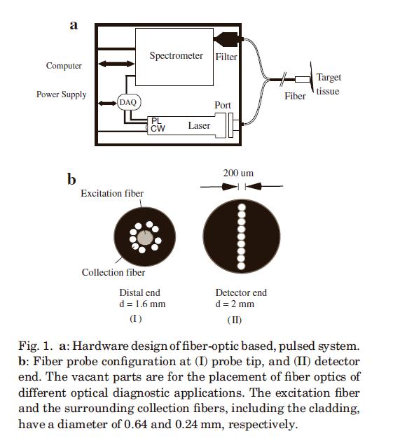
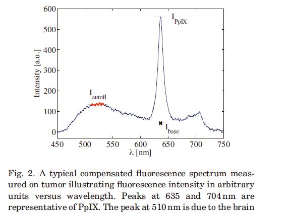
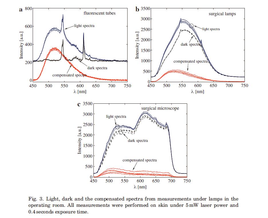
Background and Objective: Total tumor resection in patients with glioblastoma multiforme (GBM) is difficult to achieve due to the tumor’s infiltrative way of growing and morphological similarity to the surrounding functioning brain tissue. The diagnosis is usually subjectively performed using a surgical microscope. The objective of this study was to develop and evaluate a hand-held optical touch pointer using a fluorescence spectroscopy system to quantitatively distinguish healthy from malignant brain tissue intraoperatively. Study Design/Materials and Methods: A fluorescence spectroscopy system with pulsed modulation was designed considering optimum energy delivery to the tissue, minimal photobleaching of PpIX and omission of the ambient light background in the operating room (OR). 5-Aminolevulinic acid (5-ALA) of 5 mg/kg body weight was given to the patients with a presumed GBM prior to surgery. During the surgery a laser pulse at 405nm was delivered to the tissue. PpIX in glioblastoma tumor cells assigned with peaks at 635 and 704nm was detected using a fiber optical probe. Results/Conclusion: By using the pulsed fluorescence spectroscopy, PpIXfluorescence isquantitatively detected in the GBM. An effective suppression of low power lamp background from the recorded spectra in addition to a significant reduction of high power surgical lights is achieved.