
Nugzar Gomidze, Lali Kalandadze, Miranda Khajishvili, Omar Nakashidze, Izolda Jabnidze, Davit Jakobia, Kakha Makharadze
April 1, 2025 | Nugzar Gomidze, Lali Kalandadze, Miranda Khajishvili, Omar Nakashidze, Izolda Jabnidze, Davit Jakobia, Kakha Makharadze; Fluorescence spectroscopy as a novel tool in hematological diagnostics. APL Bioeng. 1 June 2025; 9 (2): 026102. https://doi.org/10.1063/5.0264155
Abstract
The present paper explores the application of fluorescence spectroscopy in erythrocyte analysis, aiming to enhance spectral diagnostics in biomedical research. The primary objective is to develop innovative methodologies for improving the precision of hematological diagnostics and disease monitoring. Utilizing 3D fluorescence spectroscopy and excitation/emission wavelength mapping, erythrocyte samples are examined across multiple wavelengths, generating distinct spectral profiles that reveal biochemical composition, oxygenation status, and metabolic alterations. Advanced data analysis enables the identification of pathological changes in erythrocytes, contributing to a more comprehensive diagnostic approach. Additionally, this study integrates fluorescence spectroscopy with traditional clinical hematological analysis, comparing spectroscopic findings with complete blood count parameters for two patients. Blood samples were subjected to fluorescence analysis under deuterium, halogen, and ultraviolet excitation sources, allowing for a detailed correlation between spectroscopic biomarkers (hemoglobin, deoxyhemoglobin, and plasma characteristics) and clinical parameters (hemoglobin concentration, hematocrit, and red blood cell indices). The findings demonstrate that fluorescence spectroscopy provides complementary diagnostic insights, detecting subtle physiological variations in blood composition that conventional methods might overlook. By integrating these two diagnostic approaches, this research highlights the potential of fluorescence-based techniques as a noninvasive and efficient tool for hematological diagnostics.
Introduction
Fluorescence spectroscopy is one of the most important techniques used across various fields, including blood analysis, biological sample examination, and other chemical and physical studies. This method is particularly notable for its ability to perform precise analysis with minimal invasiveness. In forensic science, fluorescence spectroscopy is essential for analyzing bloodstains, enhancing blood visibility, and determining its age, which is crucial for forensic investigations.1
The fundamental principles of this technique have been described in detail, illustrating how fluorescence spectroscopy operates and how it is applied in pharmaceutical sciences.2 Its application is not limited to biological samples alone; fluorescence spectroscopy is also widely used in bioanalysis, where it aids in achieving high sensitivity and specificity.3
Fluorescence-activated cell sorting (FACS) and methods for studying red blood cells are also extensively applied in molecular biology and medical research. These methods allow for the separation and analysis of cells based on fluorescence characteristics, significantly contributing to cellular research that requires identifying and analyzing various cell characteristics and functions.
The StellarNet BlackComet spectrometer is an instrument ideally suited for examining fluorescence spectra in the UV-Vis range. It can be applied to several research topics mentioned above. The BlackComet is capable of analyzing fluorescence spectra, which is valuable for studying various biological, chemical, and physical samples.
To study the microrheological properties of erythrocytes, the StellarNet BlackComet can closely examine the fluorescent characteristics of cells in a liquid medium. Additionally, this spectrometer can be used to investigate the influence of zeta potential, which is important in biological and physiological processes.
Spectrometer Schematic and Methodology
The schematic of the StellarNet BlackComet spectrometer consists of several key components that enable it to collect and analyze light spectra. The following are the main components and their functions:
The light source generates light directed at the sample under analysis. This light can be from the ultraviolet (UV), visible (Vis), or near-infrared (NIR) spectrum, depending on the application. For example, UV light might be used in biological sample analysis to induce fluorescence in the sample.
The sample is placed in a specialized holder that ensures proper transmission of light through it. The light interacting with the sample is either reflected or transmitted, allowing the spectrometer to capture its fluorescence or reflected spectrum.
The optical fiber connects the light source and the sample to the spectrometer. It transmits or reflects light from the sample to the spectrometer. The fiber can vary in length and diameter, depending on the specific research needs.
The monochromator uses a grating system to separate light into different wavelengths. This process disperses the light received from the sample into a spectrum of colors based on wavelength.
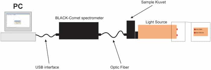
Fig. 1
The detector records the intensity of light at various wavelengths. The BlackComet spectrometer uses a CCD (charge-coupled device) detector, which is highly sensitive and allows precise signal detection across different wavelengths. The CCD detector captures the intensity data, which is then used for analysis.
The data collected by the detector are sent to a computer or data processing system for spectral analysis. Here, it is possible to perform a detailed examination of the sample’s fluorescence properties and composition (Fig. 1).