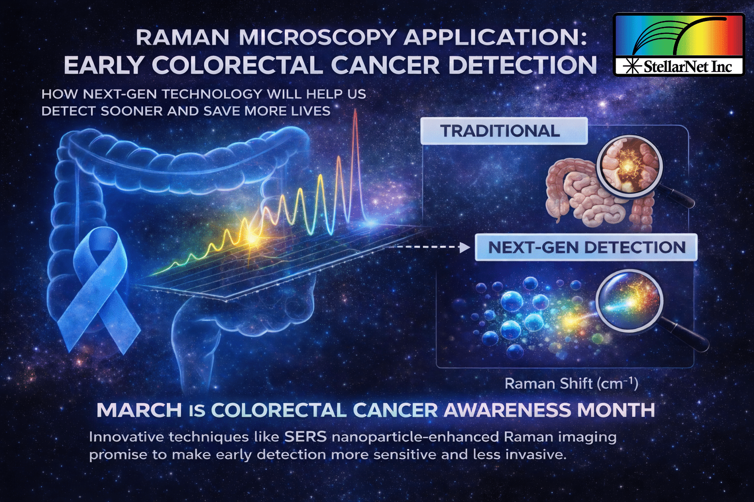
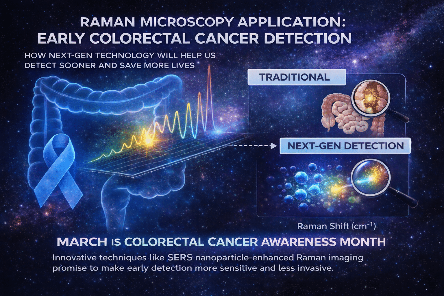
Colorectal cancer remains one of the most common malignancies worldwide, and early detection is key to improving patient outcomes. Traditional colonoscopy methods, while effective, are invasive and can miss early-stage lesions. There’s a pressing need for more sensitive, less invasive diagnostic tools.
In a recent study published in Cells (MDPI), researchers investigated how surface-enhanced Raman scattering nanoparticles (SERS NPs) could be used to detect tumor presence in a Drosophila model — highlighting the potential of Raman spectroscopy as a diagnostic modality.
Nanoparticle Uptake in Oncogenic Drosophila Midgut Measured with Surface-Enhanced Raman Spectroscopy
Maria Christou, Ayobami Fidelix, Yiorgos Apidianakis, and Chrysafis Andreou, Cells, 2024
In this study, Christou and colleagues explore how surface-enhanced Raman scattering (SERS) nanoparticles can be used as contrast agents for early tumor detection in the intestine. Using Drosophila melanogaster as a genetically controlled in vivo model, the authors demonstrate how Raman microscopy hardware combined with SERS amplification can detect disease-associated nanoparticle retention before clear morphological changes occur.
Raman & SERS Measurement Approach
The study leveraged a 785 nm Raman spectrometer system to perform whole-abdomen measurements on live flies after oral administration of silica-coated gold nanostar SERS nanoparticles. The 785 nm excitation wavelength is commonly selected in biomedical Raman applications to balance signal strength with reduced fluorescence background.
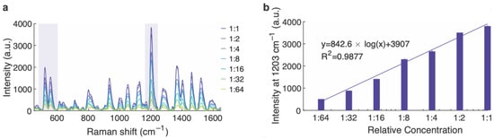
SERS nanoparticles were engineered to generate strong, distinct Raman signatures, including prominent peaks near ~1200 cm⁻¹, allowing them to be differentiated from intrinsic biological Raman signals. The plasmonic nanostar geometry enhances electromagnetic fields at sharp tips, significantly amplifying Raman scattering and improving detection sensitivity compared to conventional Raman alone.
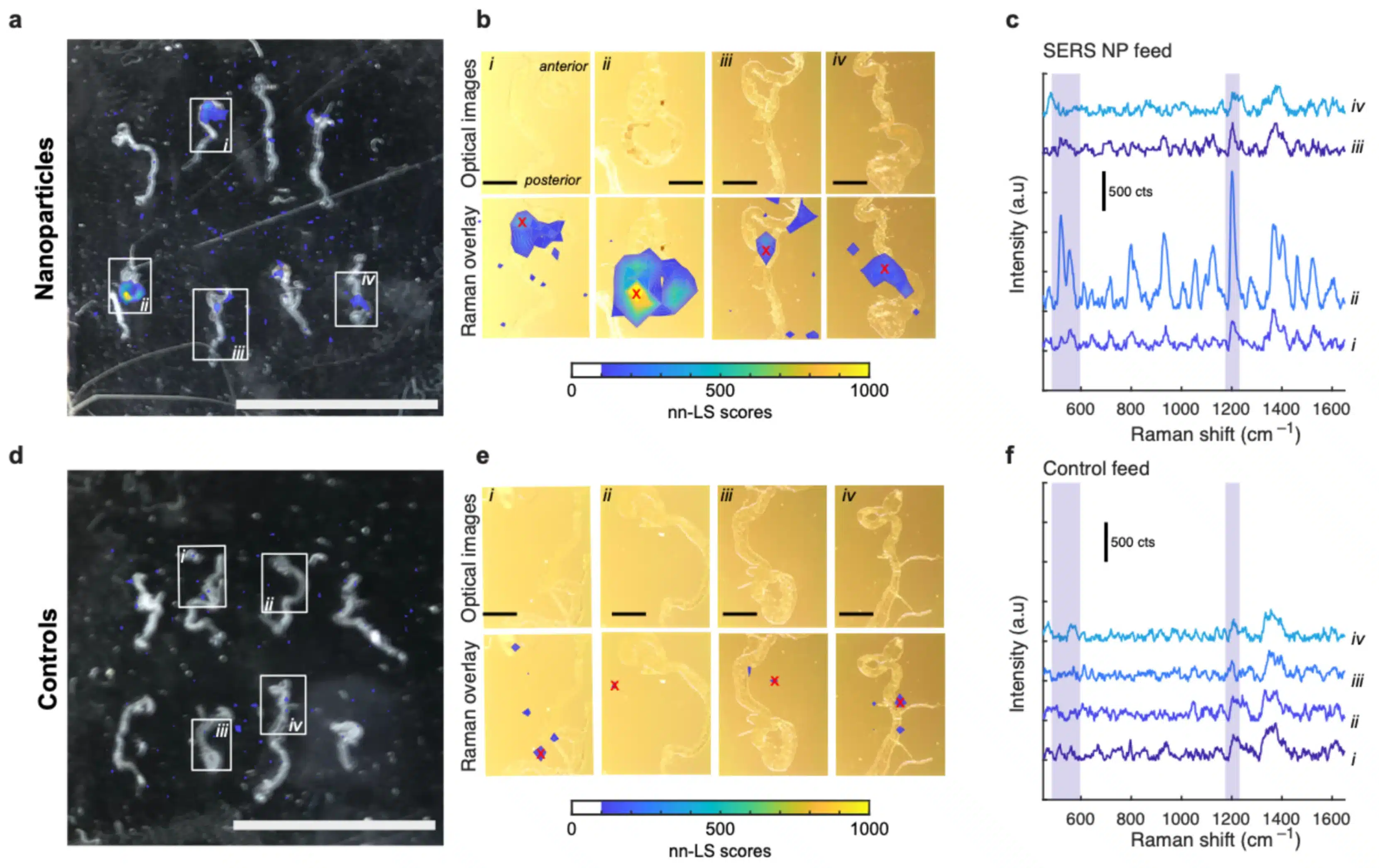
Spectra were acquired using short integration times with multiple accumulations to improve signal-to-noise performance. Standard preprocessing steps — including dark subtraction, baseline correction, and smoothing — were applied prior to analysis. To isolate nanoparticle contributions from complex biological backgrounds, the authors implemented non-negative least squares (nn-LS) regression, enabling quantitative deconvolution of overlapping spectral components.
Key Results
In healthy wild-type flies, SERS nanoparticle signals were minimal following gut clearance, suggesting limited retention in normal tissue. However, in flies genetically engineered to develop early intestinal tumors (Ras^V12 oncogenic model), statistically significant SERS signals were detected at early disease stages.
Importantly, the enhanced Raman signal was measurable before tissue disruption, indicating that SERS-based detection may reveal early tumor-associated changes not observable through traditional imaging methods. Raman mapping of dissected midguts further confirmed localized nanoparticle retention in tumor-associated regions.
Signal intensity decreased at later tumor stages, potentially due to altered feeding behavior or increased epithelial turnover — highlighting both the promise and biological complexity of in vivo SERS detection strategies.
Significant Results for Raman Microscopy
This work demonstrates the practical integration of:
-
785 nm Raman excitation
-
Plasmonic SERS nanoparticle contrast agents
-
Quantitative spectral regression analysis
-
In vivo biological measurement
Together, these components illustrate how Raman microscopy hardware combined with engineered SERS probes can detect subtle biochemical signatures associated with early disease states.
While translation to clinical colorectal cancer detection will require further optimization in delivery and background suppression, this study provides a strong proof-of-concept that Raman/SERS systems can function as highly sensitive, minimally invasive diagnostic tools.
📖 Read the full article here:
https://www.mdpi.com/2073-4409/13/16/1344