
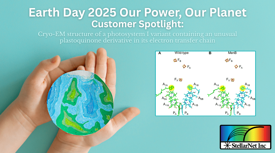
Christopher J. Gisriel, Vasily Kurashov, David F. Iwig, Brandon P. Russell, David J. Vinyard, Gary W. Brudvig, John H. Golbeck, K. V. Lakshmi
November 29, 2024 | Vol 10, Issue 48 DOI: 10.1126/sciadv.adp4937 | Yale University, The Pennsylvania State University, Louisiana State University, Rensselaer Polytechnic Institute
Abstract
Photosystem I (PS I) is a light-driven oxidoreductase responsible for converting photons into chemical bond energy. Its application for renewable energy was revolutionized by the creation of the MenB deletion (ΔmenB) variant in the cyanobacterium Synechocystis sp. PCC 6803, in which phylloquinone is replaced by plastoquinone-9 with a low binding affinity. This permits its exchange with exogenous quinones covalently coupled to dihydrogen catalysts that bind with high affinity, thereby converting PS I into a stable solar fuel catalyst. Here, we reveal the 2.03-Å-resolution cryo-EM structure of a recent MenB variant of PS I. The quinones and their binding environment are analyzed in the context of previous biophysical data, thereby enabling a protocol to solve future PS I hybrids and constructs from this genetically tractable cyanobacterium.
Introduction
Sunlight is the primary source of energy for most ecosystems, making oxygenic photosynthesis one of the most important biological processes on Earth (1). Central to oxygenic photosynthesis are the pigment-protein complexes photosystem I (PS I) (2) and photosystem II (PS II) (3) that convert the energy of light into chemical potential energy. The unusually high quantum yields of both PS I (4) and PS II (5) provide a blueprint for the design of a new generation of highly efficient artificial photosynthetic devices for the production of dihydrogen- (6) and carbon-based fuels (7–9).
The low-temperature fluorescence spectrum (fig. S2) indicates a homogeneous population of PS I complexes. The sample was subsequently negatively stained and imaged using transmission electron microscopy (fig. S3). Monodisperse, disc-shaped single particles were observed.
Two-dimensional (2D) classification and 3D reconstruction of 401 particles revealed that the particles exhibit C3 symmetry as expected. The sample was therefore considered suitable for single-particle cryo-EM. The sample was plunge frozen in liquid ethane as described in Materials and Methods. Initial cryo-EM screening also revealed monodisperse disc-shaped trimeric particles (fig. S4).
In 3D reconstruction of 599 particles, the stromal ridge of each PS I monomer could be easily distinguished. A high-resolution dataset was collected and analyzed as described in Materials and Methods (fig. S5). The final cryo-EM map exhibited a global resolution of 2.03 Å (fig. S6). Representative regions of the sharpened map are shown in Fig. 2.
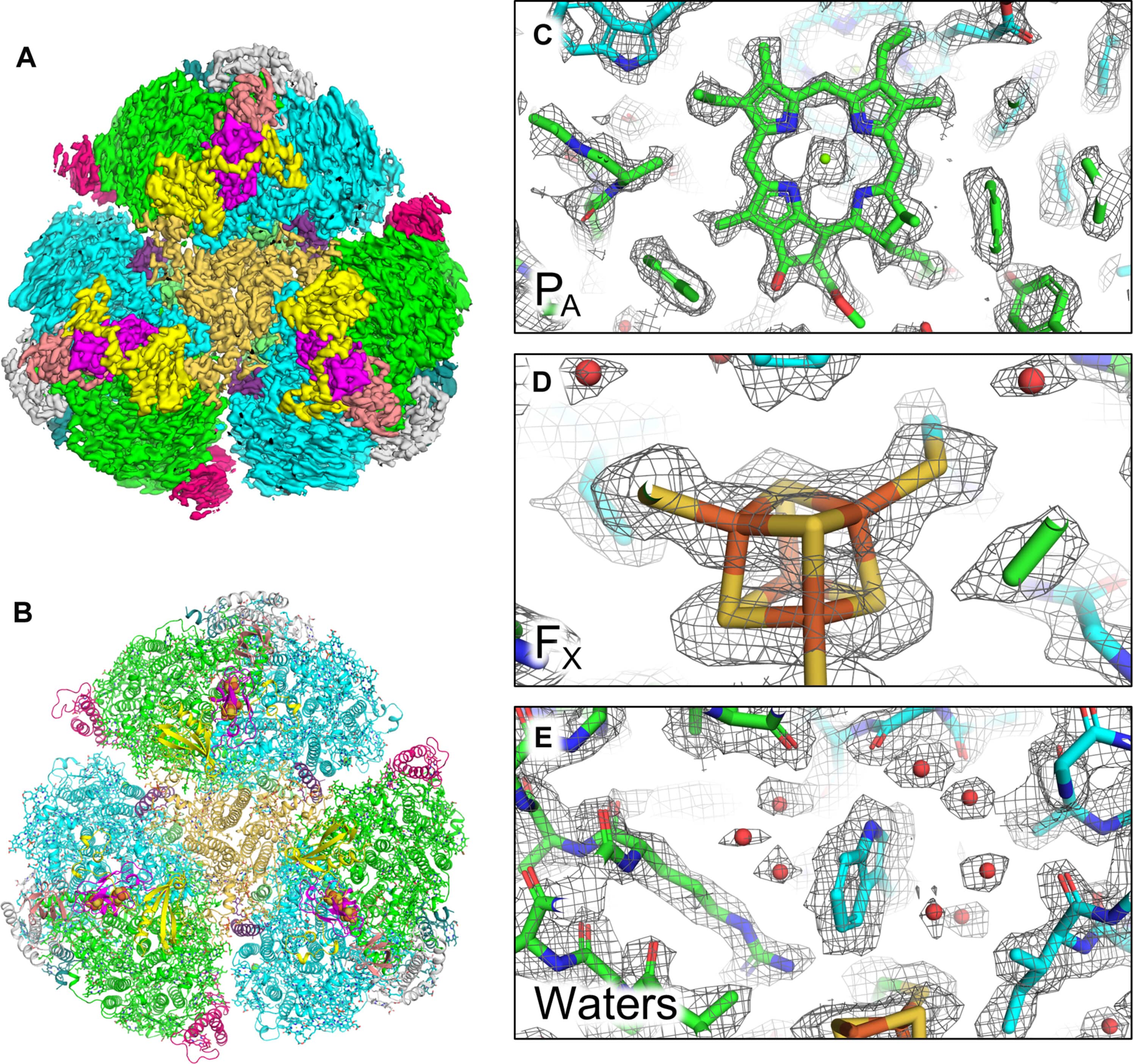
Fig. 2. Overall map and model, and representative map regions of the MenB PS I cryo-EM structure.
Low-temperature fluorescence measurements
Fluorescence was detected using a commercial spectrometer (StellarNet BLACK-Comet). To resolve any low levels of PS II contamination in the MenB PS I sample, the experiment was repeated at 80 μg Chl/ml. No fluorescence signals characteristics of PS II at ~685 or ~695 nm were observed in MenB PS I.