Y Ren, H Winter, J Rosch, K Jung, A Duross, M Landry… – ACS Applied Nano …, 2019
Abstract
Lanthanide-doped nanocrystals have been examined extensively as contrast agents for various optical molecular imaging techniques. One of the greatest strengths of these nanomaterials is their ability to enable novel imaging modalities, such as X-ray excited radioluminescence imaging, which leverages the exceptional tissue depth penetration of X-rays and reduced tissue autofluorescence. Here, we report a uniquely engineered NaGdF4/Tb@CaF2 nanoscintillator with substantial lattice mismatch through integration of coprecipitation and thermal decomposition synthetic routes. We observed greatly enhanced radioluminescence by the NaGdF4/15%Tb@CaF2 core/shell nanocrystals, which results from the minimized surface quenching and localized structure transformation. Polyethylene glycol coated NaGdF4/15%Tb@CaF2 nanocrystals demonstrated robust aqueous colloidal stability and were well tolerated by a panel of cell lines. The core/shell NaGdF4/15%Tb@CaF2 nanophosphors were subsequently decorated with targeting folate ligands and investigated as an X-ray luminescence imaging probe in vitro. Overall, the results suggest that these optimized radioluminescent nanophosphors have the potential to enable X-ray excited optical emission for biological imaging and serve as energy mediators in theranostic applications.

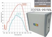
… Radioluminescence Characterization. The radioluminescence of the as-prepared β- NaGdF4:Tb core and β-NaGdF4:Tb@CaF2 core/shell nanoparticles were measured using a custom fabricated spectrometer