Skin Lesions Classification with Optical Spectroscopy – September 2010
A. Safi, V. Castaneda, T. Lasser, N. Navab- Technische Universität München, Germany
Diagnosis of benign and malign skin lesions is currently mostly relying on visual assessment and frequent biopsies performed by dermatologists. As the timely and correct diagnosis of these skin lesions is one of the most important factors in the therapeutic outcome, leveraging new technologies to assist the dermatologist seems natural. Complicating matters is a blood cancer called Cutaneous T-Cell Lymphoma, which also exhibits symptoms as skin lesions. We propose a framework using optical spectroscopy and a multi-spectral classification scheme using support vector machines to assist dermatologists in diagnosis of normal, benign and malign skin lesions. As a first step we show successful classification (94.9%) of skin lesions from regular skin in 48 patients based on 436 measurements. This forms the basis for future automated classification of different skin lesions in diseased patients.
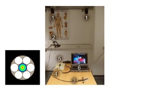
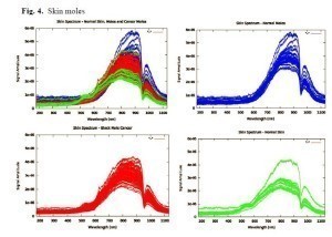
(a) Schematic of the fiber arrangement in the spectroscopy probe: 6×200μm illumination fibers arrayed around one 600μm acquisition fiber. (b) System setup: (1) tracking cameras, (2) regular camera (for augmented reality visualization), (3) tracked probe, (4) spectrometer, (5) light source, and (6) data–processing unit. The right Graph of all normalized spectra from the training data set T, color-coded as blue for skin moles, red mole cancer and green for normal skin.